research

How the interaction of physical forces and biochemical processes affects basic cell biology still eludes most scientists. We study systems-level regulation of cell and tissue biomechanics to understand its role in progression of complex diseases, and to discover new drug targets and to design precision therapeutic strategies based on mechanobiological principles. We are specifically interested in proteins that form the focal adhesion complex and actin-associated proteins that shape the mechanobiological information processing capacity of the cell. In addition to playing critical structural roles, these crosslinking and adapter proteins modulate mechanotransduction through spatial segregation of signaling proteins. We use proteomics to identify key functional proteins within the cytoskeleton or the adhesome and utilize classical cell biological as well as bioengineering methods to characterize the complex role they play in pathophysiology. We have recently showed how multiple nested network motifs-controlled expression and localization of the actin-crosslinking protein, synaptopodin, in kidney podocytes. In the past our findings have been featured as the cover story on Science Signaling. And more recently, we published a Nature Reviews Nephrology article detailing the core biophysical and biomechanical principles associated with podocyte physiology and further discuss mechanobiological pathways that could be harnessed for the discovery of podocyte-specific therapeutics.

Systems Mechanobiology

Cytoskeletal Mechanobiology

Hypertension is the second leading cause of kidney failure in the United States. It has a strong association with disease progression in both podocytopathies and polycystic kidney disease. It is still not clearly understood how biomechanical forces are mechanistically linked to cystogenesis. Terminally differentiated kidney podocytes and glomerular endothelial cells are thought to be irreversibly injured during the high biomechanical stress states imposed by hypertension. Our lab studies both tissue systems using a combination of systems biology approaches. We hypothesize that a specialized mechano-responsive actin cytoskeleton is responsible for maintaining the necessary structural integrity of podocyte foot processes. Using animal models of podocytopathies, we have identified several key proteins of the actin cytoskeleton that were previously not known to be expressed in podocytes. We use machine learning methods to integrate single-cell transcriptomics, high-content imaging and computational modeling of upstream signaling pathways to understand the dynamics of regulatory networks that control the expression of these novel gene products, and to study the spatiotemporal organization of key components within the specialized podocyte cytoskeleton. Our recent publication in Journal of American Society of Nephrology outlines the discovery of such a cytoskeletal regulator, LIM-nebulette. In our recent preprint, we identified a novel variant of ARHGEF18, a podocyte specific RhoGTPase regulator to be correlated with disease severity in DKD.

Biomechanical decision making

We explore how biomechanical properties of cells and their microenvironment impacts cellular decision making. Defects in ciliary proteins can lead to severe diseases such as polycystic kidney disease (PKD), which accounts for kidney failure in approximately 10% of all dialysis patients. Others and we have demonstrated that altered biomechanical signaling in PKD contributes to the formation of renal cysts. Utilizing a combination of murine models, in vitro studies, and computational modeling, we investigate how mutations in ciliary proteins induce changes in cell biomechanics, resulting in distinct emergent collective cell behavior. By dissecting the underlying mechanobiological mechanisms, we aim to deepen the understanding of PKD pathogenesis and identify potential unconventional therapeutic targets. Metastasis, the spread of cancer cells from the primary tumor to distant sites, is a leading cause of cancer-related mortality and depends on cancer cells' ability to deform and navigate through narrow interstitial spaces and microvasculature, as well as mobilize from the primary site. Central to these processes are cytoskeletal dynamics and mechanobiology, which regulate the motile and deformable phenotypes of cancer cells. Following up on our recent publication in Science Advances, our project aims to identify key factors that modulate these critical cellular behaviors by utilizing innovative micropatterned substrates that enable precise tracking of individual molecular dynamics during 3-D invasion. By finely tuning the dimensions of these surfaces, we mimic the mechanical challenges cancer cells encounter in the body, allowing us to dissect the mechanobiological mechanisms underlying metastasis. This approach not only advances our understanding of cancer cell behavior but also supports the development of targeted therapies to inhibit metastatic progression.

Machine learning-enabled Analytics

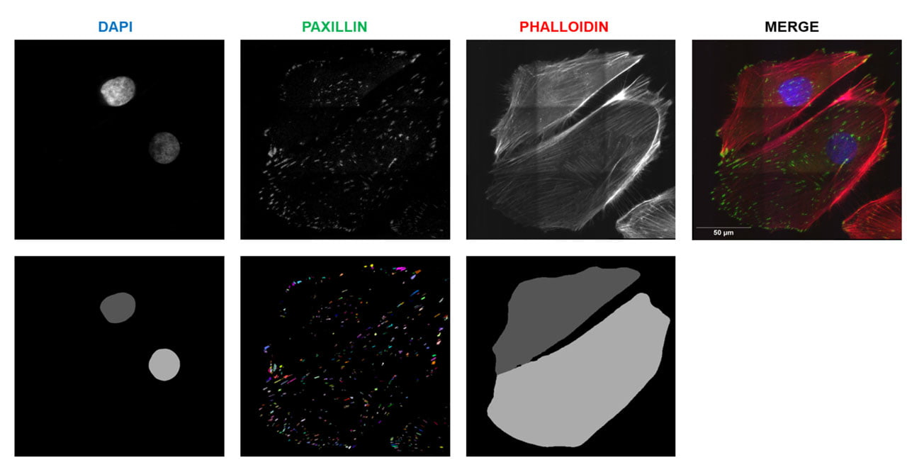
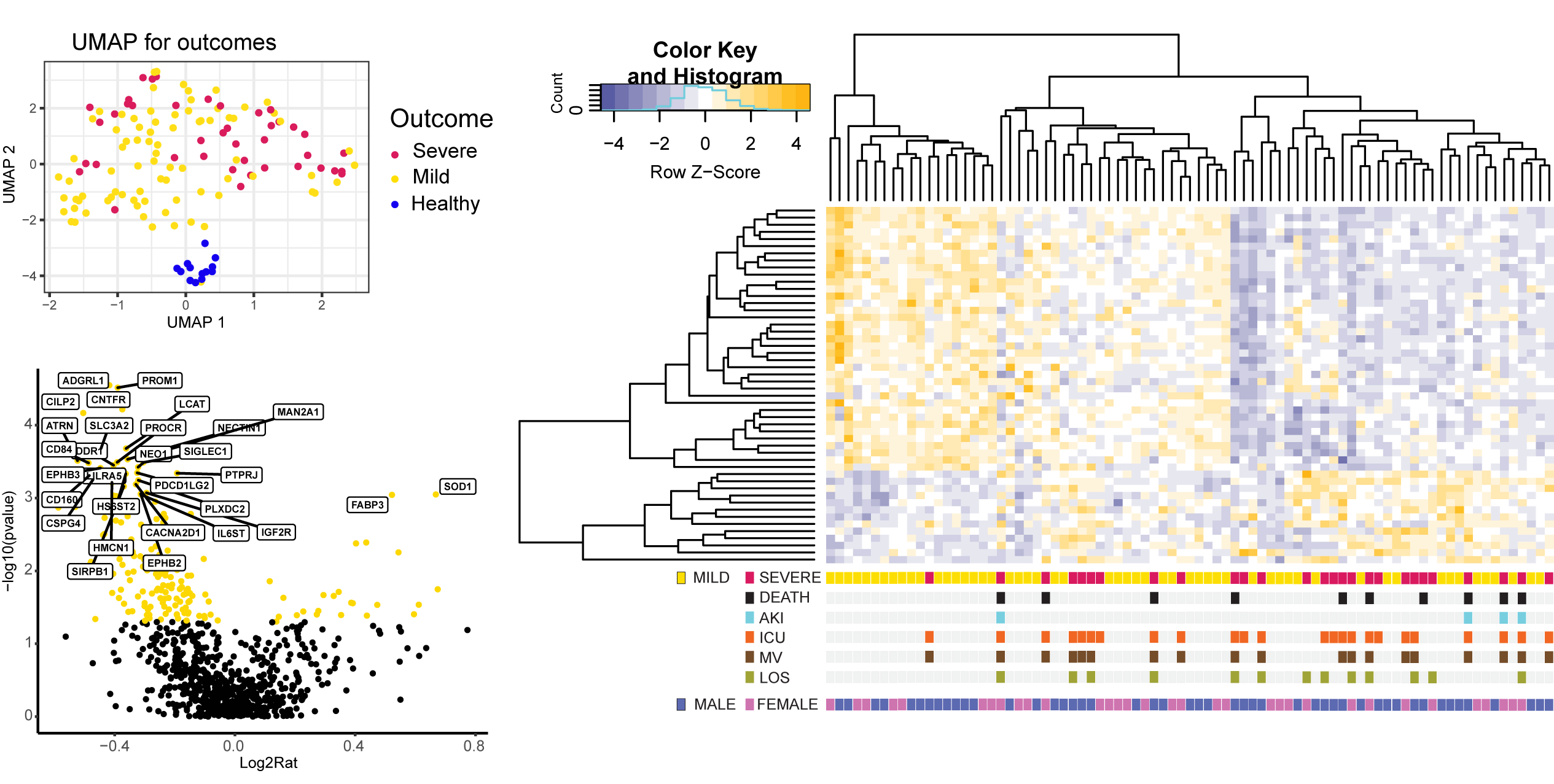
We leverage advanced machine learning (ML) techniques to enhance high-content image analysis and experimental predictions. Using neural network-based methods, we segment fluorescent images to identify cells, nuclei, mitochondria, and other subcellular structures in combination with widefield, confocal, and super-resolution microscopy. Additionally, we employ various regression models, including random forest, elastic net, and ridge regression, for diverse applications that range from predicting the effects of kinase inhibitors on cellular phenotypes to analyzing atomic force microscope elastography. We recently used machine learning to predict acute COVID-19 severity in hospitalized patients and further integrated multiomic clinical datasets to uncover mechanisms associated with COVID-19 induced kidney injury. You can read our preprint here.

ML_enabled_analytics
Covid ML

Sex Differences and Podocyte Physiology

Hypertension is an important risk factor for the development of cardiovascular diseases and is the second leading cause of end stage renal disease (ESRD) after diabetes. Interestingly, studies have reported that women are less susceptible to hypertensive kidney injury than men suggesting that sex is a potential determinant in the etiology of ESRD. Using integrated multiomics assays in a combination of in vivo models of glomerular nephropathies, we have identified a podocyte specific differential regulatory gene network that may be responsible for an increased susceptibility to early kidney damage in males compared to females. We use male and female hiPSC derived glomerular cell types to investigate the sex specific gene regulatory networks that is responsible for podocyte biomechanical resilience.

Mouse Glom
iPSC_Podocytes

Precision Therapeutics

Kidney Tissue Engineering

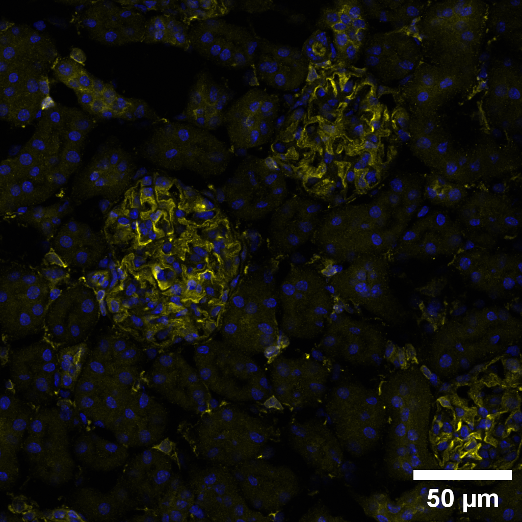
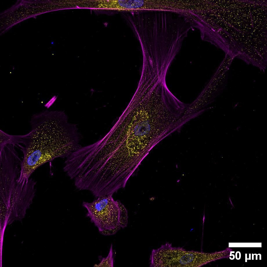
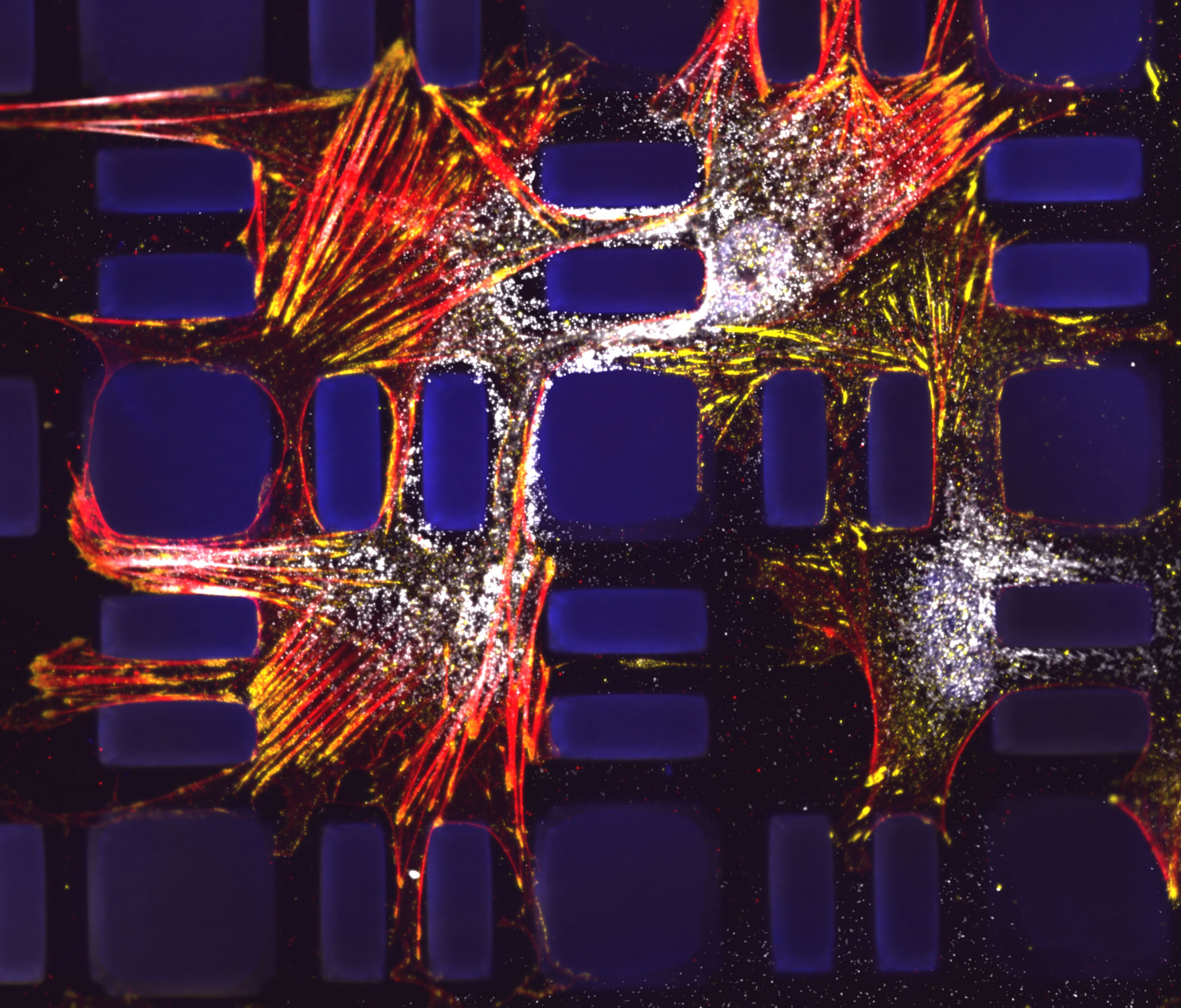
We combine nanotechnology, functional tissue engineering, and multiple-omics methodologies to study kidney biology and glomerular disease mechanisms. Despite the functional importance of their specialized morphology, isolated glomerular epithelial cells, or podocytes, do not exhibit any geometric hallmarks of their in-situ morphology. We use innovative microfabrication techniques to construct long-term culture substrates that induce morphological remodeling of kidney podocytes into arborized, or branched, shapes. This geometric remodeling leads to functional specialization of peripheral projections where filtration proteins are translocated into the periphery of the cell. This phenomenon cannot be observed in immortalized podocytes cultured on regular, or unpatterned, surfaces. This phenotypic specialization can be used as a proxy for podocyte differentiation, which enabled us to develop a kidney-on-chip platform. Using induced pluripotent stem cells (iPSCs) to generate adult human podocytes, we developed a high-content screening system that utilizes microengineered surfaces to quantitatively characterize cytoskeletal and biomechanical drug responses in kidney podocytes.

patterned_podocytes

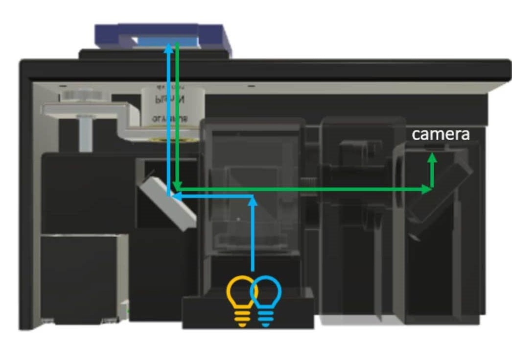
On-chip Sensor Arrays

Microphysiological systems offer a biomimetic platform for drug testing and disease modeling. While numerous advanced microphysiological systems of glomeruli have been reported, there are few robust functional assays that can be used for quantitative, rapid drug screening. In our recent ABME paper, we demonstrated the integration of a bilayer PMMA-based glomerular filtration barrier chip model with microscopic fluorescent sensor allows evaluation of barrier integrity in real-time. We are currently exploring integration of additional functional sensor systems with kidney organoids and organoid-on-chip models to build a physiologically relevant high-throughput in vitro platform.

sensor
monolayer1
monolayer2

Podocyte Kinome and Nephrotoxicity

We investigate the molecular, structural, and subcellular determinants of chemotherapy-induced podocyte damage and nephrotoxicity. Using retroactive analyses of the FDA Adverse Events Reporting System, we identify clinically used tyrosine kinase inhibitors (TKIs) that are associated with glomerular dysfunction and proteinuria. Using high-content imaging, atomic force microscope elastography, and phosphoproteomics, we identify molecular, morphological and biophysical signatures of nephrotoxicity induced by oncological TKIs. Our integrative modeling studies point to key hubs within the human kinome that are targeted by these TKIs, which then negatively impact podocyte biophysics and renal function. We are searching for potential upstream interaction nodes that can be targeted for mitigation of nephrotoxic adverse effects of oncological drugs. Our previous manuscript in Nature Communications has uncovered a direct link between cellular mechanobiology and the BCR-ABL inhibitor dasatinib. More recently, our CJASN paper , which was featured as a cover story, showed that dasatinib use in participants with chronic myeloid leukemia had a significantly higher likelihood of leading to glomerular damage evidenced by increased incidence of proteinuria compared with non-dasatinib tyrosine-kinase inhibitor treatments.

Implantable Vascular Access Technologies

Hemodialysis is the most common type of dialysis, with approximately 90% of dialysis patients undergoing this treatment. It is highly costly: notably, 1.4% of Medicare patients who have end stage kidney disease (ESKD) account for >7.2% of the overall Medicare spending. During hemodialysis treatment, blood is drawn from the patient and transferred to a dialysis machine, usually via an arteriovenous (AV) fistula. There are several complications associated with access, such as aneurysm formation, infection, damage to vessel wall, and uncontrolled bleeding. Vascular access complications limit treatment opportunities outside of the clinic, such as at home where self-cannulation would be required. To address these issues and reduce complications associated with vascular access, we developed a prototype access port that integrates well with fistulae, guides self-cannulation, and prevents aneurysms with minimal risk of back-bleeding and infections. Initial in vitro and large animal testing demonstrated that cannulation of the device is safe and simple, potentially reducing vascular damage, infection and enhancing durability.